The skin Understanding cancer Macmillan Cancer Support
(USMLE topics) Structure of the skin, layers of the epidermis, skin barrier and pigmentation. Purchase PDF (script of this video + images) here: https://www..
PPT Basic Skin Structure PowerPoint Presentation, free download ID6099891
Spend some time analyzing the skin diagram labeled above. Try to memorize the appearance and location of each structure. Learning the function of each structure will accelerate your ability to memorize, so be sure to check out our detailed article on The Integumentary System parts and functions..

Skin Structure infographic LifeMap Discovery
Hair, skin and nails Wound healing Osmosis High-Yield Notes This Osmosis High-Yield Note provides an overview of Skin Structures essentials. All Osmosis Notes are clearly laid-out and contain striking images, tables, and diagrams to help visual learners understand complex topics quickly and efficiently. Find more information about Skin Structures:
Skin diagram labeled
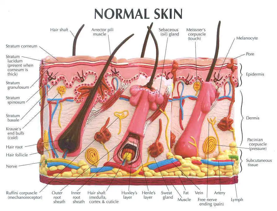
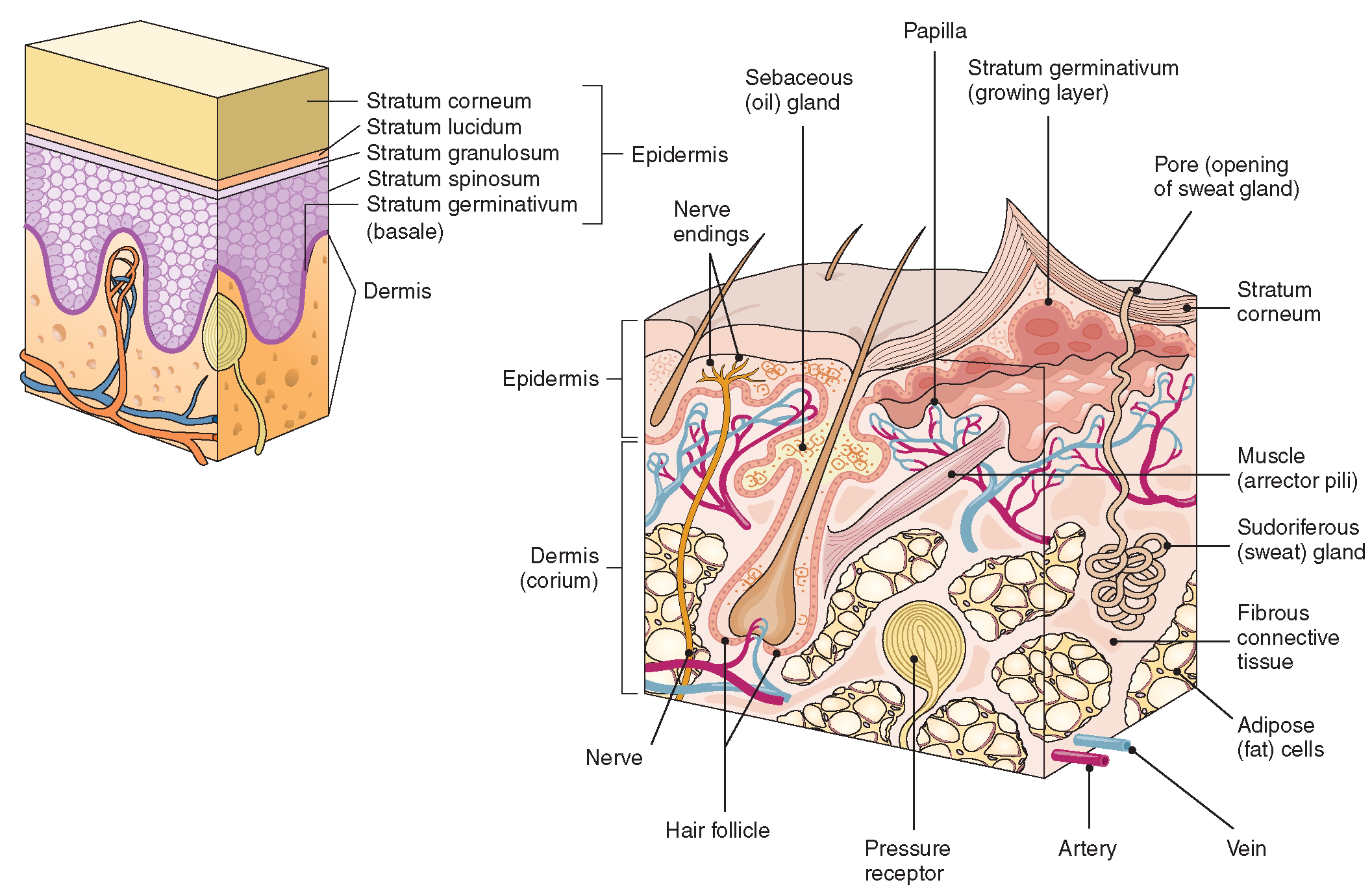
Stratum basale, also known as stratum germinativum, is the deepest layer, separated from the dermis by the basement membrane (basal lamina) and attached to the basement membrane by hemidesmosomes. The cells found in this layer are cuboidal to columnar mitotically active stem cells that are constantly producing keratinocytes.
Dermis Layers, Papillary Layer, Function Epidermis
1/3 Synonyms: none This article will describe the anatomy and histology of the skin. Undoubtedly, the skin is the largest organ in the human body; literally covering you from head to toe. The organ constitutes almost 8-20% of body mass and has a surface area of approximately 1.6 to 1.8 m2, in an adult.

Structure Of Skin Skin Structure and Function LearnFatafat
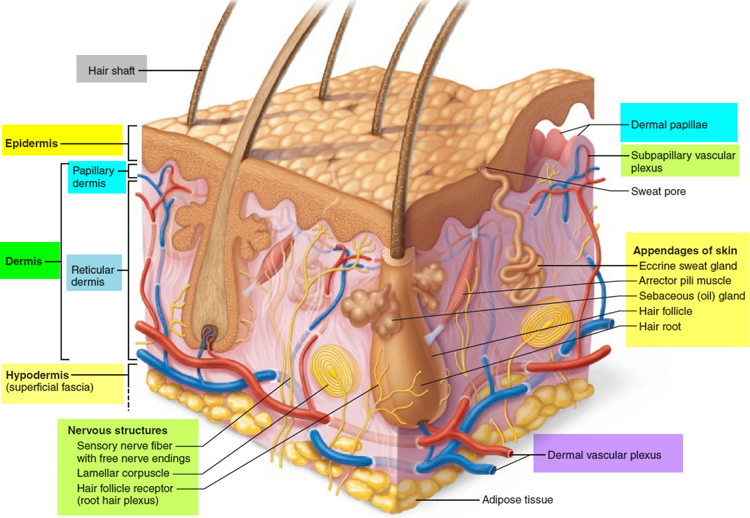
Dermis. Definition. Fibrous and elastic tissue, provides strength and elasticity to the skin and supports the epidermis, home to hair follicles, glands, nerves etc. Location. Term. Papillary Layer. Definition. Upper dermal layer, provides the epidermis with nutrients and regulates body temperature. Location.
The Integumentary System (Structure and Function) (Nursing) Part 1
Your skin continuously communicates with your brain about what is happening around you: touch, texture, temperature, tingling, pleasure, and pain. Your skin, in cooperation with your nervous.
Anatomy Of Human Skin With Labels Photograph by Hank Grebe Pixels
Diagram of human skin structure. Image. Add to collection. Rights: The University of Waikato Te Whare Wānanga o Waikato Published 1 February 2011 Size: 100 KB Referencing Hub media. The epidermis is a tough coating formed from overlapping layers of dead skin cells.

Structure of human skin cells Artofit
View this animation to learn more about layers of the skin. What are the basic functions of each of these layers? The Epidermis The epidermis is composed of keratinized, stratified squamous epithelium. It is made of four or five layers of epithelial cells, depending on its location in the body.
Schematic representation of skin structure and cell population. The... Download Scientific Diagram
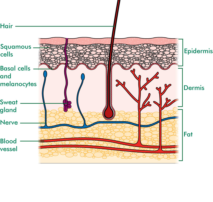
The skin is the body's largest organ. It covers the entire body. It serves as a protective shield against heat, light, injury, and infection. The skin also: Regulates body temperature. Stores water and fat. Is a sensory organ. Prevents water loss. Prevents entry of bacteria.
Layers of the Skin Anatomy and Physiology I
Acne Boils Dandruff Eczema Melanoma It is made up of the following five layers. Stratum Corneum The stratum corneum is the top layer of the epidermis. Its jobs are to: Helps your skin retain moisture Keep unwanted substances out of your body It is made of dead, flattened cells called keratinocytes that are shed approximately every two weeks.
Skin diagram to label Labelled diagram
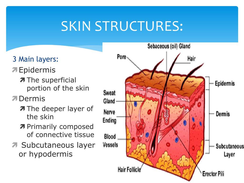
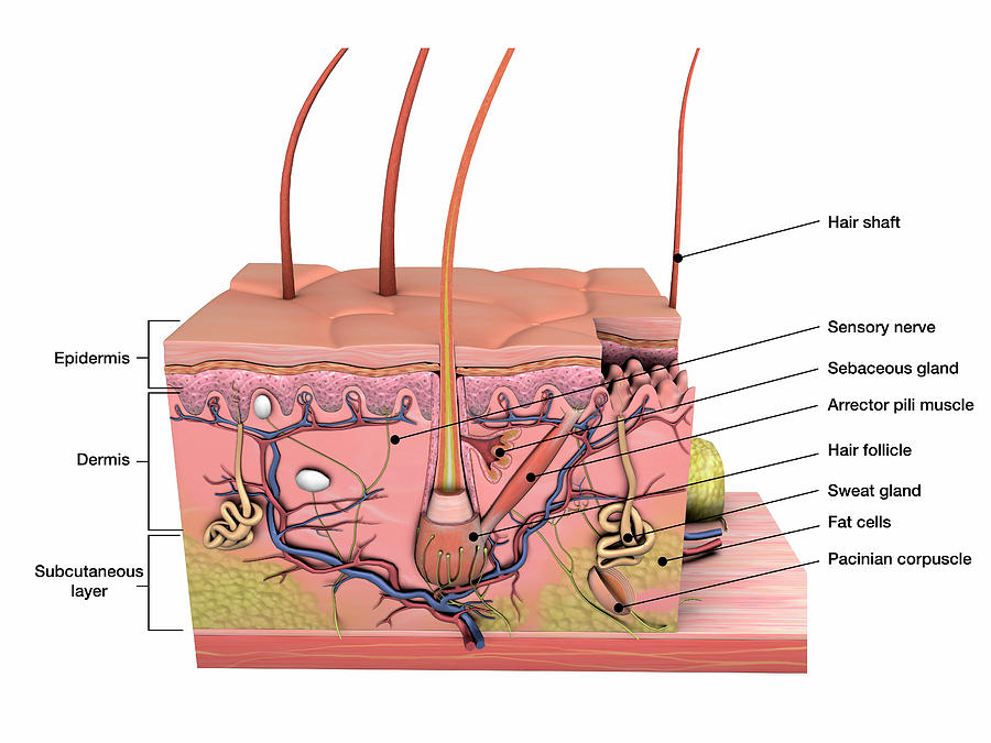
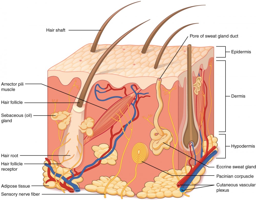
Epidermis Dermis Subcutaneous fat layer (hypodermis) Each layer has certain functions. Epidermis The epidermis is the thin outer layer of the skin. It consists of 2 primary types of cells: Keratinocytes. Keratinocytes comprise about 90% of the epidermis and are responsible for its structure and barrier functions. Melanocytes.

The Structure Of Human Skin Cells Stock Illustration Download Image Now Hair Follicle
Interactive Link The skin consists of two main layers and a closely associated layer. View this animation to learn more about layers of the skin. What are the basic functions of each of these layers? The Epidermis The epidermis is composed of keratinized, stratified squamous epithelium.
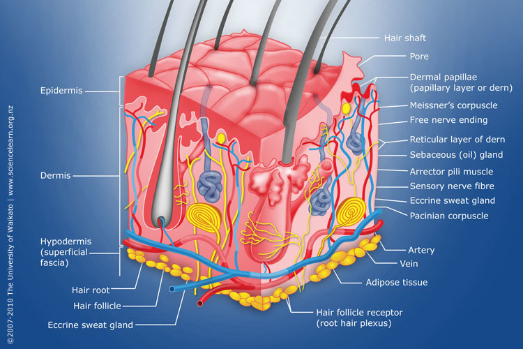
Diagram of human skin structure — Science Learning Hub
Key facts about the integumentary system; Skin: Functions: chemical and mechanical barrier, biosynthesis, control of body temperature, sensory Layers: Epidermis (Stratum Basale, Spinosum, Granulosum, Lucidum, Corneum) and dermis (papillary, reticular) Mnemonic: British and Spanish Grannies Love Cornflakes Hair: Types: vellus and terminal Structure: Follicle and bulb (shaft, inner root sheath.

loadBinary_006.gif (992×779) Skin anatomy, Integumentary system, Human integumentary system
Julia Benedetti , MD, Harvard Medical School Reviewed/Revised Dec 2021 | Modified Sep 2022 VIEW PROFESSIONAL VERSION Layers of the Skin The skin is the body's largest organ. It serves many important functions, including Protecting the body against trauma Regulating body temperature Maintaining water and electrolyte balance
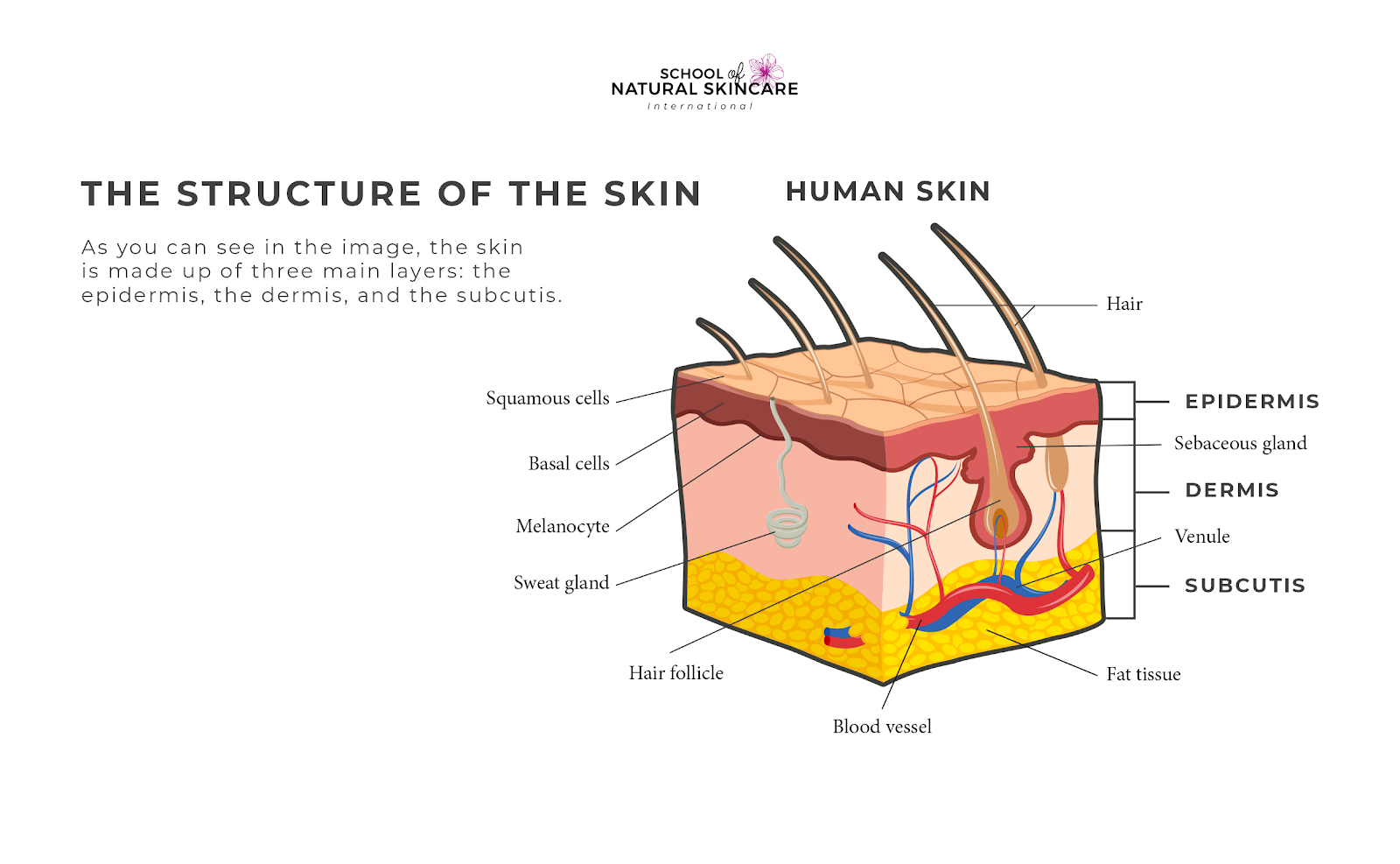
Understanding How Your Skin Works School of Natural Skincare
Figure 5.1.1 - Layers of Skin: The skin is composed of two main layers: the epidermis, made of closely packed epithelial cells, and the dermis, made of dense, irregular connective tissue that houses blood vessels, hair follicles, sweat glands, and other structures.